Contents
Scroll to:
 Vladimir A. Korolev,
Lyudmila A. Babkina,
Elena V. Felker,
Maxim A. Usachev,
Regina Y. Chertova,
Irina A. Artemova,
Diana R. Magomedova,
Vadim A. Ryzhaev
Vladimir A. Korolev,
Lyudmila A. Babkina,
Elena V. Felker,
Maxim A. Usachev,
Regina Y. Chertova,
Irina A. Artemova,
Diana R. Magomedova,
Vadim A. Ryzhaev https://doi.org/10.47470/0016-9900-2026-105-3-308-313
EDN: ndhwzy
Scroll to:
Introduction. Cadmium is one among the priority ecopollutants. The absence of a lethal or immediate impact on animals does not preclude the possibility of cadmium accumulating in specific organs and tissues, resulting in adverse consequences.
Materials and methods. Male mature Wistar rats were used for the experiment. The experimental groups received a dose of cadmium acetate dihydrate equivalent to 1/50 of the LD50, dissolved in drinking water, for 28 days, followed by 28 days on regular drinking water. The control groups consumed regular drinking water. The cadmium content was measured using the atomic adsorption method (KVANT-2AT, Russia) on days 7, 14, 21, 28, and 56 in 19 biological media.
Results. Elevated cadmium levels were observed in all studied biological media on day 7 under conditions of subchronic intoxication, except for gum and brain tissues. The highest levels of cadmium are found in the liver, small intestine, kidneys, and plasma. After the cessation of cadmium intake, the metal level is restored to the control values in the submandibular salivary glands, brain and lungs.
Limitations. Only one Wistar line was used for the laboratory experiment. The toxicant was administered orally in the form of cadmium acetate dihydrate of the same concentration.
Conclusion. The affinity of cadmium to various biological media is characterized by significant variability in the entire period of intoxication. The liver, small intestine, kidneys, and plasma act as metal accumulators. The concentration of cadmium in the liver and kidney tissues in the animal is the most likely indicator of the cadmium poisoning.
Compliance with ethical standards. The study was approved by the Regional Ethics Committee of Kursk State Medical University (protocol No. 3 of October 17, 2022), carried out in accordance with the ethical principles of working with animals for scientific purposes governed by «Directive 2010/63/EU of the European Parliament and of the Council on the protection of animals used for scientific purposes» and national standards. This article does not contain results from studies involving human subjects.
Contribution:
Korolev V.A. – the concept and design of the study, editing;
Babkina L.A. – the concept and design of the study, information search, statistical processing and analysis of research results, writing text;
Felker E.V. – the concept and design of the study, analysis of the results;
Usachev M.A. – conducting an experiment, analyzing samples, and processing data;
Chertova R.Y. – information search, experiment, data processing;
Artemova I.A – conducting an experiment, analyzing samples;
Magomedova D.R., Ryzhaev V.A. – conducting an experiment, collecting data.
All authors are responsible for the integrity of all parts of the manuscript and approval of the manuscript final version.
Conflict of interest. The authors declare no conflict of interest.
Funding. The work was carried out at the expense of Kursk State Medical University.
Received: June 20, 2025 / Accepted: October 15, 2025 / Published: April 17, 2026
Korolev V.A., Babkina L.A., Felker E.V., Usachev M.A., Chertova R.Y., Artemova I.A., Magomedova D.R., Ryzhaev V.A. Features of cadmium distribution in the body under conditions of subchronic intoxication. Hygiene and Sanitation. 2026;105(3):308-313. https://doi.org/10.47470/0016-9900-2026-105-3-308-313. EDN: ndhwzy
Introduction
Biogeochemical changes to the environment resulting from human activity lead to the accumulation of heavy metals in soil, atmospheric air, water and plants [1–3]. Cadmium is one of the priority environmental pollutants. The main routes of cadmium entry into the human body are linked to migration through food chains, from the atmosphere via breathing, and during smoking [1, 4]. Within the body, cadmium is then redistributed to various organs, followed by detoxification and accumulation [1, 5]. A long biological half-life, combined with high toxicity and the absence of physiological elimination mechanisms, can pose risks to public health [6].
The adverse effects of cadmium affect virtually all human organs and systems [1, 7, 8]; the patterns of accumulation depend on species, sex, age, and the physiological condition of the body [9]. A deficiency or excess of certain elements in the diet can lead to increased cadmium absorption [10]. The effects of cadmium in the body are determined by the route of exposure, the dose, and the duration of exposure [11]. High doses lead to cell apoptosis, whereas subchronic and chronic exposure activate adaptive mechanisms in response to the external environment [6, 12]. The distribution of Cd across organs varies depending on the dose and the specific compound entering the body [10]. Accumulation in biological systems depends on the presence of effective cellular transport mechanisms and ligands (thiol groups, metallothioneins, albumins, etc.) [10]. Studies have examined the effects of Cd on the main target organs of model animals – the liver and kidneys [13]. However, there is insufficient information on the specific dynamics of cadmium accumulation in target organs during the intoxication period, as well as in other biological compartments. Cadmium chloride is predominantly used as the toxicant [13]. Oral administration of Cd reflects the natural route of its entry into the human body as an environmental pollutant and takes into account the specific absorption characteristics of various organs.
Studying the dynamics of cadmium distribution in model animals during subchronic exposure will identify the most vulnerable organs and systems, with the aim of developing optimal prevention strategies to reduce the risk of adverse health effects, and will also aid in the development of new approaches to monitoring and controlling the concentration of this element in biological systems.
The aim of the study was to investigate the dynamics of cadmium levels in various organs of model animals following subchronic oral exposure.
Materials and methods
The experiment was conducted at the Experimental Biology Clinic of the Kursk State Medical University (KGMU), a Federal State Budgetary Educational Institution of Higher Education under the Ministry of Health of Russia. Eight-week-old male Wistar rats weighing 220–230 g were used. The animals were housed in individual cages under standard conditions.
Access to food and drinking water was unrestricted. For 28 days, the experimental rats received cadmium acetate dihydrate (CAS No. 5743-04-4, 99.5% purity, Russia) at a dose of 1/50 LD₅₀ (LD₅₀ = 360 mg/kg). Over the following 28 days, the animals were switched to normal drinking water. The control groups of rats received normal drinking water throughout the study period. Each group of animals consisted of 10 individuals. The concentration of cadmium in tap water and feed was below the limit of detection. Animals were removed from the experiment on days 7, 14, 21, 28 and 56 by euthanasia under ether anesthesia.
Cadmium distribution was analyzed in oral fluid, submandibular salivary glands, gingival tissue, hard periodontal tissues, the small and large intestines, the liver, plasma, red blood cell mass, submandibular lymph nodes, spleen, lungs, kidneys, testes, heart, sternum, brain, and fur. Unstimulated oral fluid (V = 50 µl) was collected from live rats using a JoanLab pipette dispenser (DPAOP-1-10-100 JoanLab, ‘Lenpipet Thermo Scientific’, Russia), and centrifuged for 20 minutes at 1500 rpm (rotor F-45-12-11; MiniSpin, Germany). Blood was collected from the right ventricle of the heart and centrifuged for 15 minutes (3000 rpm, rotor F-45-12-11; MiniSpin, Germany) at +4 °C to obtain plasma and red blood cell pellet. Fur was collected from the area of the animal’s back below the projection of the shoulder blades (S = 1 cm²) using medical scissors. It was then degreased with acetone. Organs were removed, rinsed with saline solution and used to prepare homogenates. The cadmium content in the samples was determined in the mineralised extract, obtained by atomic absorption spectrometry (‘KVANT-2AT’, Russia) in absorption mode using LT-6M spectral lamps (λ = 228.8 nm), and expressed in µg/ml or µg/g. Atomisation was carried out in an acetylene–air flame. Statistical analysis of the results was performed using Statistica 13.0 (StatSoft, USA). The Shapiro–Wilk test was used to determine the distribution type of cadmium content in organs, and the Mann–Whitney test was used to compare independent samples. Statistical significance was set at p < 0.05.
Results
Cadmium concentrations in biological media are presented in the table. Cd concentrations in various tissues and organs of control animals were negligible and varied: the highest levels were found in the kidneys (0.459–0.486 µg/g), the tissues of the large intestine (0.37–0.395 µg/g) and erythrocyte mass (0.326–0.366 µg/ml), and the lowest in the heart (0.042–0.045 µg/g), plasma (0.051–0.06 µg/ml) and submandibular lymph nodes (0.076–0.08 µg/g).
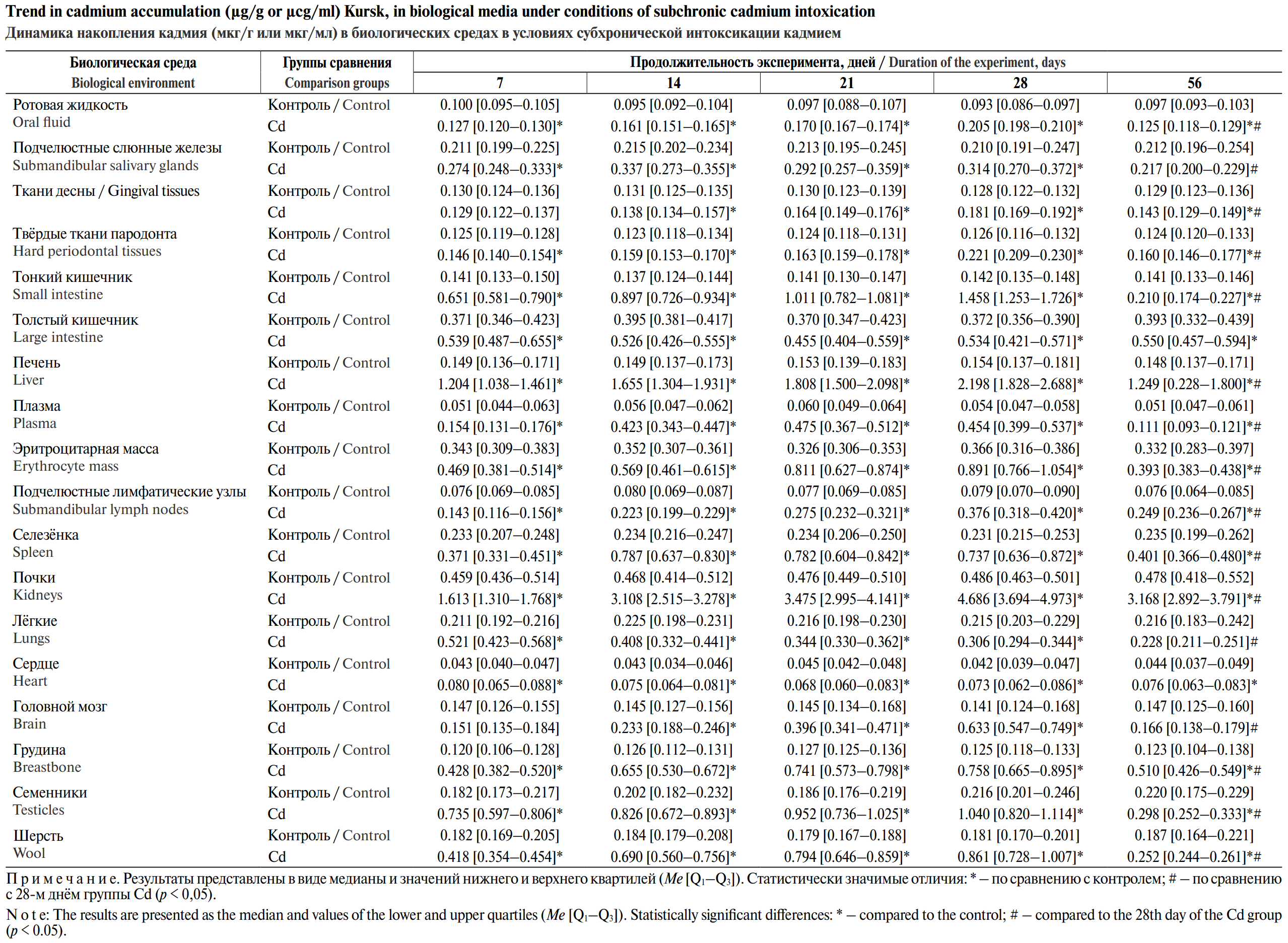
The administration of cadmium at a dose of 1/50 LD₅₀ via drinking water led to an increase in its concentration compared with the control in all biological media studied. At the same time, specificity was observed in the dynamics of Cd accumulation in different tissues and organs. Elevated levels were observed after 7 days of exposure in all tissue samples and organs studied (p < 0.05), with the exception of gingival tissue (p = 0.9097) and brain tissue (p = 0.3075). Cd levels increased significantly in the liver (8.1-fold), small intestine tissues (4.64-fold), testes (4.11-fold), sternum (3.58-fold), kidneys (3.5-fold), plasma (3-fold), lungs (2.48-fold) and wool (2.33-fold). After 14 days of exposure, cadmium concentrations were found to be higher than in the control group in all biological samples examined, with the highest increases observed in the liver (11.11-fold), plasma (7.55-fold), small intestine wall (6.55-fold) and kidneys (6.64-fold). In the lungs, a decrease in cadmium levels was observed compared with day 7: with levels 1.81 times higher than the control. On the 21st day of intoxication, the greatest increase was observed in the liver (11.82-fold), plasma (7.92-fold), kidneys (7.3-fold) and the small intestine wall (7.17-fold). The liver, small intestine, kidneys and blood plasma are greatest accumulators of cadmium on the 28th day of intoxication as well. At the same time, a redistribution of accumulation levels is noted: the content in the liver increases by 14.27 times, in the small intestine by 10.27 times, in the kidneys by 9.64 times and in plasma by 8.41 times.
Based on an analysis of the dynamics of cadmium content during subchronic intoxication, groups of tissues and organs can be identified according to the specificity of the element’s accumulation (see figure).
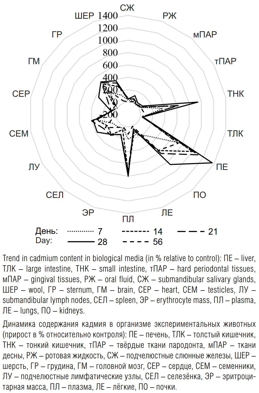
In most tissues and organs, a continuous increase in cadmium concentration was observed throughout the experiment, peaking on the 28th day of intoxication: the liver (by 1367%), the small intestine (by 943%), kidneys (by 857%), plasma (by 800%), submandibular lymph nodes (by 376%), brain (by 350%), oral fluid (by 122.2%), hard periodontal tissues (by 69.23%), gingival tissues (by 41.41%). For tissues of the large intestine and heart, a slight variation in elevated cadmium levels was characteristic over the 28-day period of intoxication: by 24.32–45.95% for the large intestine, and by 75–86.5% for the heart. In the lungs, the maximum increase in cadmium concentration was recorded on the 7th day of intoxication (by 148%); subsequently, a decrease occurred, and by the 28th day the increase was 47.62%. In the submandibular salivary glands, the most significant increase in cadmium content was observed on the 14th day of intoxication (by 61.9%). In the tissues of the spleen and sternum, the maximum increase was detected on the 14th and 28th days of intoxication: by 222–239% in the spleen and by 442–485% in the sternum. The maximum increase in Cd content on the 21st day, with levels remaining high until the end of intoxication, was observed in the erythrocytes (by 145–147%), testes (by 373–400%) and fur (by 339–378%) of rats.
Based on an analysis of the percentage change in cadmium content in biological media relative to the control, the following groups of organs and tissues can be identified: 1) with marked accumulation (by more than 500%) – liver, small intestine, kidneys and plasma; 2) with moderate accumulation (more than 300%) – sternum, brain, testes, submandibular lymph nodes, fur; 3) with negligible accumulation (less than 300%) – oral fluid, submandibular salivary glands, gingival tissue, large intestine, red blood cells, spleen, heart, lungs.
Once cadmium administration was discontinued, cadmium levels returned to levels indistinguishable from those in control animals in the submandibular salivary glands (p = 0.4274), the brain (p = 0.1041) and the lungs (p = 0.1405). In the large intestine wall and heart, following the recovery period, cadmium levels remained at the level observed on the 28th day of intoxication (p= 0.162 and p = 0.7338, respectively). In the other biological compartments studied, cadmium levels decreased after the cessation of additional cadmium exposure but didn’t reach the values of the control group.
Discussion
Prolonged intake of small doses of cadmium via drinking water is accompanied by its selective accumulation in various organs and tissues of animals [14]. Variability in cadmium accumulation by biological substrates may be due to the characteristics of their anatomical structure, physiological processes, mechanisms of element transport and maintenance of homeostasis, and processes of xenobiotic biotransformation [15].
Upon oral administration, cadmium is absorbed predominantly in the duodenum and the proximal jejunum [16]. The highest concentration in enterocytes of all types of cellular transporters (ABC transporters, ZIP, DMT1, TPP transporters) [16] involved in cadmium transport may explain its significant accumulation in the small intestinal wall throughout the entire period of intoxication and the reduction in its concentration to almost the level of control animals after cessation of external intake. Cadmium readily forms complexes with thiol-containing biomolecules and, in the form of a conjugate of these ligands, is also absorbed by enterocytes. The more pronounced increase in cadmium content on the 7th day of intoxication in the small intestinal wall of rats compared with plasma and kidneys may be due to dose-dependent absorption. At low doses, most of Cd is localized in the intestinal epithelium, but as intake increases, the cells’ ability to retain Cd decreases, and most of it penetrates the epithelial membrane and is absorbed by other organs [17]. The good regenerative capacity of epithelial cells may explain the greater ability of enterocytes to restore cadmium homeostasis compared with other target organs.
High plasma cadmium levels throughout the entire period of intoxication may be due to its release from enterocytes into the portal blood via basolateral ABC transporter proteins in the form of complexes with various ligands (metallothionein, beta-2-microglobulin, albumin, immunoglobulin G, lipocalin- 2, glutathione) [15]. Residual cadmium in plasma is capable of being taken up by erythrocytes [15, 17], which may explain its higher concentration in blood cells compared with plasma in both control and experimental animals, as well as the more pronounced increase only on the 21st day of intoxication.
Metallothioneins play a significant role in the distribution of cadmium to individual tissues and organs via the bloodstream [13, 19]. Actively absorbed by hepatocytes via the cellular transporter system in the form of conjugates with biomolecules, Cd induces the synthesis of metallothioneins and accumulates in the liver in a bound form [16]. The ability of the ‘cadmium–metallothionein’ complex to be filtered in the renal glomeruli and reabsorbed in the cells of the proximal tubules leads to the stimulation of metallothionein synthesis in the kidneys and, consequently, to an increase in cadmium concentration [19]. The liver and kidneys are the primary targets and storage sites for cadmium when it is ingested via drinking water and food [17, 20]. Thus, significant accumulation of Cd in the liver and kidneys throughout the entire period of intoxication and a slow decrease in concentration after cessation of exposure may have been due to the induction of metallothionein synthesis followed by Cd-MT complex formation [18]. A more pronounced increase in cadmium levels in the liver compared with the kidneys indicated chronic exposure to low levels of the element, whereas the reverse ratio indicated acute effects of high doses [14, 17].
Prolonged oral exposure to cadmium inhibits calcium absorption in the intestine and contributes to bone degeneration [6, 17]. The replacement of calcium by cadmium in bone tissue may explain the five-fold higher Cd content in the sternum of experimental rats compared with the control group from the 14th day of exposure and its slight decrease after cessation of metal intake. However, less pronounced accumulation of Cd was observed in the hard tissues of the periodontium, which may be associated with more developed elimination systems.
The testes may act as a critical organ in male rats with chronic cadmium intoxication [1, 16, 17, 21, 22]. The uptake of cadmium into testicular cells is associated with cadmium mimicking zinc in cellular transporters and the formation of thiol conjugates [16]. Maximum accumulation as early as the 21st day of exposure, followed by the maintenance of high levels in the subsequent days, was possibly due to the saturation of zinc transporters [16].
The immunomodulatory or immunosuppressive effects of cadmium may be due to cadmium’s ability to accumulate in lymph node cells [23].
The toxic effects of cadmium in various organs and tissues are due to their capacity to accumulate cadmium, the intensity of metabolic processes, and the presence of a transporter system that facilitates the uptake of Cd into cells and its redistribution within the body.
Conclusion
Prolonged exposure to potentially toxic substances in quantities insufficient to cause death or acute effects may lead to adverse effects. Cadmium’s affinity for various biological media is characterized by significant variability throughout the entire period of intoxication. For most tissues and organs, cadmium levels increased with additional exposure throughout the entire period of intoxication, with the liver, small intestine, kidneys and plasma acting as reservoirs. In the lungs and salivary glands, the maximum increase in cadmium levels was observed in the early stages of intoxication, followed by a decrease. In the tissues of the large intestine and heart, Cd concentrations remained virtually unchanged throughout the period of exposure. Cessation of exposure led to a return of Cd levels to baseline values in the submandibular salivary glands, brain and lungs; to levels remaining at those of the final day of intoxication in the large intestine wall and heart; and to a decrease without reaching baseline values in all other biological compartments.
The most likely indicator of cadmium intoxication in an animal’s body is the concentration in liver tissue, whilst the content in the kidneys and the ratio of metal in the liver to that in the kidneys serve as additional indicators reflecting the level of exposure.
1. Davidova S., Milushev V., Satchanska G. The mechanisms of cadmium toxicity in living organisms. Toxics. 2024; 12(12): 875. https://doi.org/10.3390/toxics12120875
2. Babkina L.A., Lukyanchikov D.S., Lukyanchikova O.V. Peculiarities of heavy metals accumulation by dooryard plantain’s leaves (Plantago major L.) in the urbanized territories. Samarskii nauchnyi vestnik. 2018; 7(1): 19–24. https://elibrary.ru/yrhzsy (in Russian)
3. Nevedrov N.P. Profile distribution and reserves of heavy metals in soils of different genesis in the cities of the western part of the Central Chernozem region. Agrofizika. 2024; (4): 9–15. https://doi.org/10.25695/agrph.2024.04.02 https://elibrary.ru/kqykbr (in Russian)
4. Lech T., Sadlik J.K. Cadmium concentration in human autopsy tissues. Biol. Trace Elem. Res. 2017; 179(2): 172–7. https://doi.org/10.1007/s12011-017-0959-5
5. Apiamu A., Asagba S.O., Tonukari N.J. Role of Anthocleista vogelii in serum antioxidant defence system in cadmium-induced oxidative stress in Wistar rats. Beni-Suef Univ. J. Basic Appl. Sci. 2019; 8(1): 12. https://doi.org/10.1186/s43088-019-0012-1
6. Charkiewicz A.E., Omeljaniuk W.J., Nowak K., Garley M., Nikliński J. Cadmium toxicity and health effects – a brief summary. Molecules. 2023; 28(18): 6620. https://doi.org/10.3390/molecules28186620
7. Rani A., Kumar A., Lal A., Pant M. Cellular mechanisms of cadmium-induced toxicity: a review. Int. J. Environ. Health Res. 2014; 24(4): 378–99. https://doi.org/10.1080/09603123.2013.835032
8. Flora S.J.S. Metal poisoning: threat and management. J. Med. Sci. 2009; 2(2): 4–26.
9. Blazka M.E., Nolan C.V., Shaikh Z.A. Developmental and sex differences in cadmium distribution and metallothionein induction and localization. J. Appl. Toxicol. 1988; 8(3): 217–22. https://doi.org/10.1002/jat.2550080310
10. Swiergosz-Kowalewska R. Cadmium distribution and toxicity in tissues of small rodents. Microsc. Res. Tech. 2001; 55(3): 208–22. https://doi.org/10.1002/jemt.1171
11. Neathery M.W., Miller W.J. Metabolism and toxicity of cadmium, mercury, and lead in animals: a review. J. Dairy Sci. 1975; 58(12): 1767–81. https://doi.org/10.3168/jds.S0022-0302(75)84785-0
12. Skipper A., Sims J.N., Yedjou C.G., Tchounwou P.B. Cadmium chloride induces DNA damage and apoptosis of human liver carcinoma cells via oxidative stress. Int. J. Environ. Res. Public Health. 2016; 13(1): 88. https://doi.org/10.3390/ijerph13010088
13. Gizatullina A.A., Valova Ya.V., Smolyankin D.A., Khusnutdinova N.Yu., Karimov D.O., Karimov D.D., et al. Effect of chronic intake of cadmium chloride on the transcriptional activity of metallothionein and zinc transporter genes. Gigiena i Sanitaria (Hygiene and Sanitation, Russian journal). 2024; 103(2): 158–64. https://doi.org/10.47470/0016-9900-2024-103-2-158-164 https://elibrary.ru/qtnvci (in Russian)
14. Ping Z., Bi Z., Huanping L., An L.Z. Heavy metal concentrations in five tissues of chickens from a mining area. Pol. J. Environ. Stud. 2014; 23(6): 2375–9.
15. Cygan-Szczegielniak D. The levels of mineral elements and toxic metals in the longissimus lumborum muscle, hair and selected organs of red deer (Cervus elaphus L.) in Poland. Animals. 2021; 11(5): 1231. https://doi.org/10.3390/ani11051231
16. Bridges C.C., Zalups R.K. Molecular and ionic mimicry and the transport of toxic metals. Toxicol. Appl. Pharmacol. 2005; 204(3): 274–308. https://doi.org/10.1016/j.taap.2004.09.007
17. Scheuhammer A.M. The chronic toxicity of aluminium, cadmium, mercury, and lead in birds: a review. Environ. Pollut. 1987; 46(4): 263–95. https://doi.org/10.1016/0269-7491(87)90173-4
18. Nordberg G.F., Nogawa K., Nordberg M., Friberg L.T. Cadmium. In: Handbook on the Toxicology of Metals. Academic Press; 2007: 445–86. https://doi.org/10.1016/b978-012369413-3/50078-1
19. Linšak D.T., Linšak Z., Spirić Z., Srebočan E., Glad M., Cenov A., et al. Influence of cadmium on metallothione in expression and products of lipid peroxidation in the organs of hares (Lepus europaeus Pallas). J. Appl. Toxicol. 2014; 34(3): 289–95. https://doi.org/10.1002/jat.2880
20. Carpen E., Andreani G., Isani G. Trace elements in unconventional animals: a 40-year experience. J. Trace Elem. Med. Biol. 2017; 43: 169–179. https://doi.org/10.1016/j.jtemb.2017.02.003
21. Usmanova E.N., Karimov D.O., Daukaev R.A., Khusnutdinova N.Yu., Afonkina S.R., Musabirov D.E., et al. Peculiarities of cadmium accumulation in the gonads in heterosexual laboratory animals. Gigiena i Sanitaria (Hygiene and Sanitation, Russian journal). 2024; 103(5): 483–88. https://doi.org/10.47470/0016-9900-2024-103-5-483-488 https://elibrary.ru/qdnqjt (in Russian)
22. Bhardwaj J.K., Panchal H., Saraf P. Cadmium as a testicular toxicant: a review. J. Appl. Toxicol. 2021; 41(1): 105–17. https://doi.org/10.1002/jat.4055
23. Wang Z., Sun Y., Yao W., Ba Q., Wang H. Effects of cadmium exposure on the immune system and immunoregulation. Front. Immunol. 2021; 12: 695484. https://doi.org/10.3389/fimmu.2021.695484
DSc (Biology) sciences, professor, head, Department of biology, medical genetics, and ecology, Kursk State Medical University, Kursk, 305041, Russian Federation
e-mail: medecol1@yandex.ru
PhD (Biology), associate professor, Department of biology, medical genetics, and ecology, Kursk State Medical University, Kursk, 305041, Russian Federation
e-mail: L-Babkina@yandex.ru
PhD (Medicine), associate professor, Department of surgical dentistry and maxillofacial surgery, Kursk State Medical University, Kursk, 305041, Russian Federation
e-mail: felkerev@kursksmu.net
PhD student, Department of biology, medical genetics, and ecology, Kursk State Medical University, Kursk, 305041, Russian Federation
e-mail: usachev.macsim@yandex.ru
PhD student, assistant lecturer, Department of biology, medical genetics, and ecology, Kursk State Medical University, Kursk, 305041, Russian Federation
e-mail: chertovary@kursksmu.net
Assistant lecturer, Department of microbiology, virology, and immunology Kursk State Medical University, Kursk, 305041, Russian Federation
e-mail: artemovaia@kursksmu.net
Assistant lecturer, Department of biology, medical genetics, and ecology, Kursk State Medical University, Kursk, 305041, Russian Federation
e-mail: magomedovadr@kursksmu.net
5th year student, Pediatric Faculty, Kursk State Medical University, Kursk, 305041, Russian Federation
e-mail: ryzhaev.vadim.2016@yandex.ru
Korolev V.A., Babkina L.A., Felker E.V., Usachev M.A., Chertova R.Y., Artemova I.A., Magomedova D.R., Ryzhaev V.A. Features of cadmium distribution in the body under conditions of subchronic intoxication. Hygiene and Sanitation. 2026;105(3):308-313. https://doi.org/10.47470/0016-9900-2026-105-3-308-313. EDN: ndhwzy
Мы в ВК:

 Email this article (Login required)
Email the author (Login required)
Email this article (Login required)
Email the author (Login required) 141014, The Moscow region, Mytishchi, 2, Semashko street
Federal Scientific Center of Hygiene named after F.F. Erisman
Phone: +7 916 885 28 02
e-mail: gigsan@list.ru