Contents
Scroll to:
https://doi.org/10.47470/0016-9900-2026-105-5-486-492
EDN: slpiqn
Scroll to:
Introduction. The inherent limitations of conventional water quality and safety monitoring for public drinking water supplies underscore the urgent need for “in vitro” cellular models. Such models provide a robust framework for the rapid screening and identification of chemical contaminants in water samples.
The purpose of the work is to substantiate, based on the results of experimental modeling, the possibility of using the HuTu 80 human cell culture to detect chemical contamination of water sources for household and drinking water use by the population.
Materials and methods. The quality and safety of drinking water sources were assessed using an expanded set of forty five sanitary-chemical indicators, including the mandatory minimum (MR 2.1.4.0176–20), in compliance with the regulatory standards established in SanPiN 1.2.3685–21. The response of the HuTu 80 cell line to water samples was assessed by monitoring changes in mitochondrial dehydrogenase activity following a 48-hour exposure period.
Results. 5 out of 10 water samples exerted an adverse impact on the HuTu 80 cell line, although only two samples showed concentrations of chemical substances (magnesium, sodium, lithium ions, as well as chlorides and sulfates) exceeding established regulatory limits. Experimental modeling of the native water composition for the first time demonstrated that metabolic inhibition could be attributed to the presence of magnesium, sulfate, and lithium ions at concentrations both above and below their respective maximum permissible levels. Furthermore, the combined effect of these inorganic ions induced a more pronounced shift in metabolic activity compared to the influence of each individual component.
Limitations. The present study is limited by the requirement for specialized equipment and precision analytical instruments, a relatively small sample size, and the necessity to account for microbiological factors.
Conclusions. The results of the study on native water samples and experimental modeling of their composition demonstrated the potential of using the HuTu 80 cell culture to detect chemical pollution in drinking water sources when maximum permissible concentrations for individual sanitary-chemical indicators are exceeded.
Compliance with ethical standards. Study approval was provided by the Local Ethics Committee of the Saratov Hygiene Medical Research Center of the FBSI «FSC Medical and Preventive Health Risk Management Technologies (meeting protocol No. 18 dated SSeptember 1 2022) and was conducted in accordance with the generally accepted scientific principles of the Declaration of Helsinki of the World Medical Association (2013 revision).
Contribution:
Kuzyanov D.A. – concept and design of the study, material collection, statistical data processing, manuscript writing;
Moiseeva E.M. – material collection and processing, manuscript writing;
Mikerov A.N. – editing, approval of the final manuscript version;
Erdniev L.P. – editing;
Lutcevich I.N. – editing, approval of the final manuscript version.
All authors are responsible for the integrity of all parts of the manuscript and approval of the manuscript final version.
Conflict of interest. The authors declare no conflict of interest.
Funding. The study had no sponsorship.
Received: March 24, 2026 / Accepted: May 20, 2026 / Published: June 18, 2026
Kuzyanov D.A., Moiseeva E.M., Mikerov A.N., Erdniev L.P., Lutsevich I.N. Justification of the prospects of using HuTu 80 cell culture for detecting chemical contamination of water sources for household and drinking water use by the population. Hygiene and Sanitation. 2026;105(5):486-492. https://doi.org/10.47470/0016-9900-2026-105-5-486-492. EDN: slpiqn
Introduction
The assessment of the quality and safety of water from sources used for household and drinking water supply (hereinafter referred to as ‘water sources’) in the Russian Federation involves carrying out laboratory tests, including those for the quantitative determination of chemical substances in accordance with the requirements of regulatory and methodological documents (SanPiN 1.2.3685–21¹, MR 2.1.4.0176–20², etc.). However, this approach does not fully account for the likelihood of adverse effects arising from the combined exposure to chemical substances and their transformation products, as well as the presence of chemical substances not included in the list of monitored parameters (MR 2.1.4.0176–20²) [1]. Consequently, the potential risks to public health associated with exposure to chemical factors in the living environment may be considerably higher, necessitating the supplementation the existing approach to the hygienic assessment of the quality of the human living environment with new developments. One promising avenue for addressing this challenge is the use of alternative biological models, including in vitro cell-based test systems. A key advantage of these research methods is that testing is carried out directly on human cell cultures [2].
To detect chemical contamination in water sources used for household and drinking water supply, it may be advisable to supplement traditional chemical-analytical methods with approaches based on the use of biological systems, such as cell cultures of the human gastrointestinal tract [2–4]. The HuTu 80 human duodenal adenocarcinoma cell line is frequently used to study the mechanisms underlying the development of toxic effects associated with exposure to chemical compounds [5–7]. HuTu 80 cells are epithelial in nature and represent stable pathological clones of duodenal surface cells [5, 8, 9]. The HuTu 80 cell line is effective in assessing microbial contamination of water and could potentially be used to complement physicochemical analytical methods when evaluating water quality according to sanitary-chemical parameters [10, 11].
The aim of the study is to justify the use of the HuTu 80 human cell culture for detecting chemical contamination in water sources used for household and drinking water supply, based on the results of experimental modelling.
Materials and methods
The study utilised water samples from sources intended for household and drinking water supply (n = 10), collected in 2024–2025 in the Saratov and Astrakhan regions, and the Republic of Kalmykia. Sampling, transport and storage of the samples were carried out in accordance with the requirements of GOST 31861–2012³. Sanitary and chemical analyses of the water samples were conducted at the Laboratory of Physicochemical
Analysis of the Saratov Regional Centre for Hygiene, part of the Federal State Budgetary Institution ‘Federal Research Centre for Medical and Preventive Technologies for Public Health Risk Management’ of Rospotrebnadzor, using titrimetric and spectrophotometric methods of analysis, as well as atomic absorption spectroscopy. Water quality was assessed against an extended list of parameters (45 physicochemical parameters), including the mandatory minimum (MR 2.1.4.0176–202), in accordance with the regulatory values established by SanPiN 1.2.3685–2¹.
To simulate the chemical composition of native water samples, aqueous salt solutions were used with calculated mass concentrations of chloride ions (NaCl and CaCl2), sulphate ions (Na2SO4), magnesium ions (MgSO4·5H2O) and lithium ions (Li2SO4). Pure analytical-grade reagents (Vekton JSC, St Petersburg) were used in this study.” The HuTu 80 human duodenal adenocarcinoma monolayer cell line, which can be passaged, was used as a test system to assess the effects of exposure to water samples and model solutions (the culture was obtained from the “Vertebrate Cell Culture Collection” shared-use centre at the Institute of Cytology, Russian Academy of Sciences, St Petersburg). HuTu 80 cells were cultured in complete DMEM medium (PanEco, Russia) containing 1% essential amino acids, 2 mM glutamine, 1 mM sodium pyruvate, 1.5 g/L sodium bicarbonate, 4.5 g/L D-glucose, and 10% bovine foetal serum (Capricorn, Germany), with the addition of penicillin/streptomycin (100 units/mL each, Capricorn, Germany). Cultivation conditions were as follows: temperature of 37 °C in an atmosphere of 5% CO2 in a culture flask (NEST Biotechnology Co., LTD., Wuxi, China), using a thermostat (Binder GmbH, Germany). Cells that had undergone no more than 10 passages were used in the experiments [12].
Water samples (2 ml) were concentrated by incubation for 1.5 hours in a water bath (LOIp, Russia) at 70 °C. This treatment method allowed the samples to be concentrated, whilst simultaneously inactivating the vegetative forms of microorganisms, which is necessary for working with cell cultures in vitro. The volume of the concentrated samples was 25% of the original (four-fold concentration). After cooling the sample, meant for analysis, to room temperature, its volume was restored to the original level using complete DMEM medium. To assess the effectiveness of water sample decontamination, a control plating of each sample under investigation was carried out on LB (Luria Bertani) [13] in Petri dishes in duplicate and incubated for five days in a thermostat at +37 °C.
The effects of the water samples on the HuTu 80 cell culture were assessed using a test based on the inhibition of cell proliferation, measured by the activity of mitochondrial dehydrogenases that reduce tetrazolium derivatives (MTT assay). Once 70–90% confluence was reached, intact cells were harvested from the culture flask using a trypsin-EDTA solution (0.25% trypsin and 0.2% EDTA, Capricorn, Germany) and seeded into 96-well culture plates (NEST Biotechnology Co., LTD., Wuxi, China) at a seeding density of 1 ∙ 10⁴ cells per well. The cells were incubated in the plates until a 70 per cent monolayer had formed over a period of 48 hours, after which the medium was removed and the wells were filled with water samples prepared according to the method described above (eight wells for the analysis of a single water sample). As control samples, the wells containing cells were filled with 200 µl of culture medium comprising 25% distilled water and 75% complete DMEM medium.
The model pollutants were dissolved in DMEM medium, sterilised by filtration through a syringe filter with a pore size of 0.1 µm, and 50 µl of each was added to wells containing 150 µl of complete DMEM medium.
Cells cultured in medium based on the prepared water samples or model pollutant solutions were incubated for 48 hours at 37 °C in an atmosphere a 5% СО2. At the end of the incubation period, the medium was removed and 100 µl of MTT solution (3-(4,5-dimethylthiazol-2-yl)-2,5-diphenyltetrazolium bromide, Sisco Research Laboratory Pvt. Ltd., India) to a concentration of 1 mg/ml, and the cells were incubated for three hours under the same conditions. The MTT solution was removed, and the formazan crystals formed in the viable cells were dissolved in 200 µl of 99% dimethyl sulphoxide (DMSO, Molekula Ltd., UK) and incubated for 15 minutes under the same conditions.
The MTT assay values for both experimental and control replicates were determined as the difference between the mean optical density values of the eluate, measured at 530 and 620 nm using a microplate reader (Pikon, Russia).
Cell viability was calculated using the formula (1):
Viability = (ODo / ODc) ∙ 100%, (1)
where ODo – the difference in optical density of the eluate from the experimental group at wavelengths of 530 and 620 nm; ODc – the difference in optical density of the eluate from the control group at wavelengths of 530 and 620 nm.
The effects of water samples on the HuTu 80 cell culture were assessed based on a statistically significant (p < 0.05) change in their respiratory activity following a 48-hour incubation with the test water sample compared with the control (cell respiratory activity after a 48-hour incubation in complete medium was taken as 100%).
Statistical analysis of the data was performed using the Statistica for Windows v. 7.0 software package (StatSoft Inc) and Microsoft Excel 2010. Mean values and standard deviations were calculated; a two-tailed Student’s t-test was used, as well as one-way analysis of variance (ANOVA) employing Fisher’s F-test, followed by Tukey’s multiple comparison test at a significance level of p < 0.05. The effective concentrations of inorganic ions (EC20) and their 95% confidence intervals were determined by probit analysis using the EPA Benchmark Dose Software (BMDS, version 3.3, US Environmental Protection Agency).
Results
Physicochemical analysis of water samples from water sources revealed that five samples exceeded regulatory limits for chemical content. The range of exceedances of public health standards for sanitary-chemical parameters in the samples studied ranged from 1 to 2.7 times the maximum permissible concentration (MAC). In parallel with the Physicochemical analysis, the effects of the water samples on the HuTu 80 human duodenal adenocarcinoma cell culture were assessed. The results are presented in Table 1.
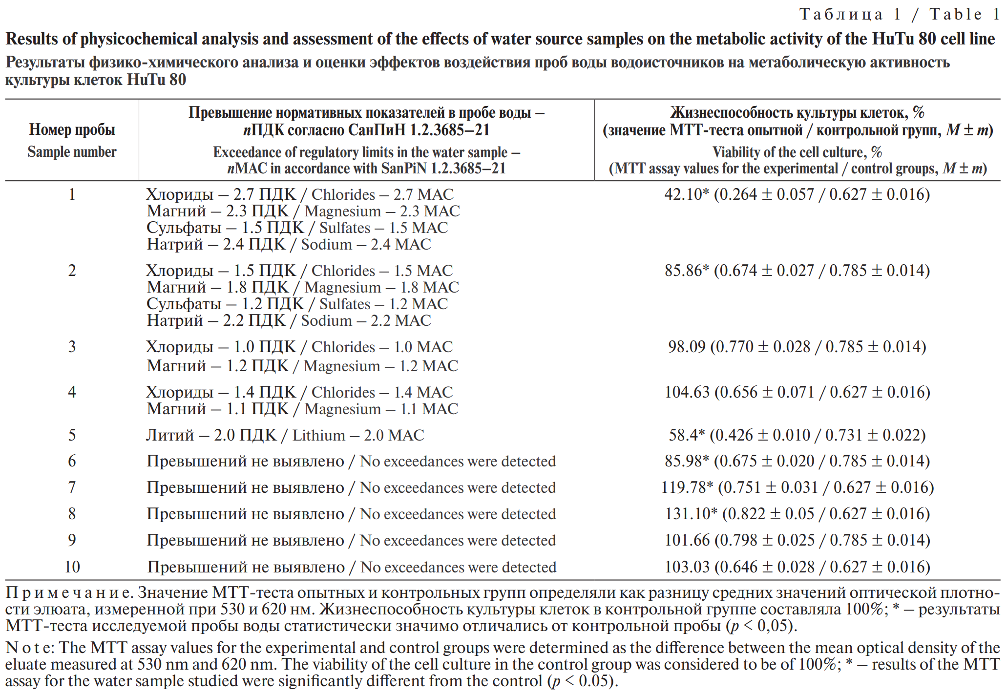
According to the results presented in Table 1, the six water samples under investigation had an effect on the metabolic activity of the HuTu 80 cell culture following 48-hour exposure. Water samples Nos. 1, 2 and 5 induced a cytotoxic effect, manifested as a statistically significant reduction in the metabolic activity of the cells by 57.9 per cent, 14.1 per cent, and 36.2 per cent, respectively, compared with the control (100 per cent) at p < 0.05. The levels in these water samples exceeded the hygienic standards by a factor of between 1.5 and 2.7 times the maximum permissible concentration (MAC) for chlorides, between 1.8 and 2.3 times the MAC for magnesium, between 1.2 and 1.5 times the MAC for sulphates, and between 2.2 and 2.4 times the MAC for sodium, 2 MAC for lithium.
Exceedances of the public health standards for certain chemical substances in water samples Nos. 3 and 4 (up to 1.4 MAC for chloride and magnesium ions) were not accompanied by any adverse effects; however, water samples Nos. 6, 7 and 8 caused a statistically significant change in the metabolic activity of the cells compared with the control (p < 0.05), despite the absence of any exceedances of the health standards for chemical content (see Table 1).
To establish a link between the presence of chemical substances at concentrations exceeding hygienic standards and changes in the metabolic activity of cell cultures upon exposure to them, the composition of the water samples under investigation was replicated in model experiments. The chemical composition of the model solutions included substances identified in this study as priority water pollutants in water sources (lithium, sodium and magnesium ions, as well as chloride and sulphate ions) at concentrations corresponding to the native water samples Nos. 1, 2, 3, 4 and 5. The results are presented in.
Exposure to the model solutions and their corresponding native samples resulted in a statistically significant reduction in the metabolic activity of the cells compared with the control (p < 0.01). Differences in the percentage change in the viability of the HuTu 80 cell culture following exposure to native water samples Nos. 2, 3, 4 and 5 and their corresponding model solutions were not statistically significant (p > 0.05). However, the metabolic activity of the cells following exposure to native sample No. 1 and its model solution differed statistically significantly (the difference in the effects of exposure was 42.7%) (Table 2).
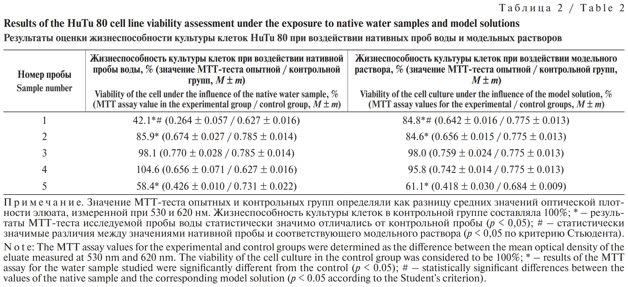
Model salt solutions, whose ionic composition corresponded to water samples No. 3 and No. 4 (exceeding the MAC by up to 1.4 times), did not cause a statistically significant reduction in cell metabolic activity (viability 98.1% and 104.6% respectively; p > 0.05) compared with the control, which is consistent with the data obtained when assessing the effects of the corresponding native samples (p < 0.05) (see Table 2).
To establish a relationship between the concentrations of magnesium, lithium and sodium ions, as well as chloride and sulphate ions in water, and their effect on the metabolic activity of the HuTu 80 cell line, the effective concentrations (EC20) of the ions were determined (Table 3).
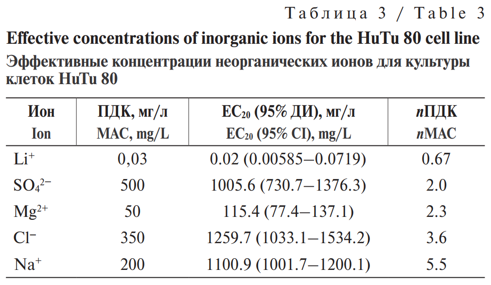
It has been established that the calculated EC20 values for magnesium, sodium, chloride and sulphate ions exceed the established hygienic standards for their concentration in water (up to 5.5 MAC) (see Table 3). At the same time, the HuTu 80 cell culture was found to be highly sensitive to lithium ions, the sensitivity manifesting at concentrations below the maximum permissible levels.
To assess the combined effect of chemical substances on the metabolic activity of the HuTu 80 cell culture, the effects of individual compounds were determined at concentrations corresponding to sample No. 1, characterised by the highest number of parameters exceeding the maximum permissible values. During the study, solutions of MgSO4, Na2SO4, CaCl2 and NaCl were prepared with ion concentrations corresponding to those in sample No. 1. The effect of the salt solutions on the metabolic activity of the cells was assessed by comparing them with one another, as well as with a model solution and a control (without toxicants) (Table 4).
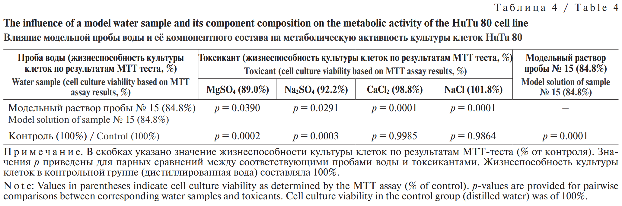
The results of a one-way analysis of variance (ANOVA) showed that the ions present in the salts of model solution No. 1 (Mg²+, Ca²+, Na+, Cl−, SO4²−), affected the metabolic activity of the cells to varying degrees compared with the control. A statistically significant change in cell metabolism was observed following exposure to the sulphate-containing salts MgSO4 (p = 0.039) and Na2SO4 (p = 0.0291). Furthermore, the combined effect of the components of model sample No. 1 caused the most pronounced reduction in culture viability (p = 0.0001).
Discussion
The presence of chemical substances in water, intended for household and drinking purposes, at concentrations exceeding regulatory limits poses a potential threat to public health and compromises the hygienic conditions of water use. Consequently, there is growing interest in the development and application of methods based on the use of biological models in an in vitro system as markers of water quality deterioration according to sanitary-chemical indicators [14, 15]. With the aim of determining the potential for using the HuTu 80 human duodenal cell culture to detect chemical contamination in water samples from sources used for household and drinking water supply, we conducted, for the first time, a study of native water samples and carried out experimental modelling of the effects of the identified pollutants (magnesium, sodium and lithium ions, as well as chloride and sulphate ions).
A comparison of the results of the assessment of the effects of the native water samples on the HuTu 80 cell culture with their chemical composition made it possible to establish a link between changes in cellular metabolic activity and the concentration of certain ions in the samples. Thus, a reduction in cellular metabolic activity was observed in response to exposure to water samples in which the levels of chlorides (up to 2.7 MAC), magnesium (up to 2.3 MAC), sulphates (up to 1.5 MAC), sodium (up to 2.4 MAC) and lithium (up to 2 MAC). The results obtained are corroborated by data from experimental modelling of the effects of salt solutions (matching the composition of the native samples), according to which there were no statistically significant differences in changes in the metabolic activity of the cell culture between the native samples and the model variants (p > 0.05).
It is important to note that concentrations of magnesium ions and chloride ions exceeding regulatory limits by up to 1.4 times the MAC did not lead to any change in metabolic activity at the cellular level. Thus, it has been confirmed that these chemical substances are low-toxic and are regulated on the basis of organoleptic indicators of harmfulness. The adverse effects of magnesium, sodium, lithium, chloride and sulphate ions in those samples, where they were observed, are most likely attributable to osmotic stress, disruption of ionic homeostasis and DNA fragmentation caused by their excessive concentration in the culture medium [16–19].
A water sample from a water source is a complex, multi-component solution in which chemical substances are present in various concentrations and combinations. According to the results of modelling of the combined effect of priority water pollutants from water sources on the HuTu 80 cell culture, it was found that the effect of the mixture of substances (model sample) was statistically significantly greater than their individual effects. The most pronounced change in metabolic activity was recorded when the cell culture was exposed to native water sample No. 1 (cell viability 42 per cent) compared with the model solution (cell viability 84.8 per cent); this is due to the combined action of all the chemicals contained in the water sample, as well as, probably, the presence of pollutants not included in the list of those assessed during the chemical-analytical study.
To determine the sensitivity of the cell culture to the concentration of priority pollutants in the water, an assessment of their effective concentrations was carried out. It was shown that the sensitivity of the cell culture to sodium, magnesium, sulphate and chloride ions, at a 20% change in metabolic activity, corresponds to concentrations of these substances ranging from 2 to 5.5 times the maximum permissible concentration (MAC). Sensitivity to lithium ions was observed at concentrations below the maximum permissible level, demonstrating the potential for using this cell culture in monitoring studies and for detecting chemical contamination of water sources by these substances.
The process of identification of substances, not included in the mandatory list of parameters monitored in water from water sources within supervisory measures, requires the extended laboratory tests of water quality. According to MR 2.1.4.0176–202, a comprehensive assessment of water source using an extended list of parameters must be carried out at least once every five years.
However, in the emergency circumstances of the arrival into the water source of chemical substances, including previously unidentified ones, resulting from anthropogenic pollution, the HuTu 80 cell culture could potentially be used as a screening test for the rapid detection of chemical contamination in water. Thus, the observed change in the metabolic activity of HuTu 80 cells following exposure to water samples from water sources, in which no exceedances of regulatory limits were recorded, may potentially indicate the presence of chemicals not included in the list of regulated substances. Thus, the test may serve as a basis for conducting an extended chemical-analytical investigation of the water source with the aim of identifying specific compounds to which the cell cultures are known to be sensitive, based on the results of laboratory tests.
The absence of a specific reaction in human cell cultures to the chemical composition of the test water sample, and the lack of a well-founded mechanism for extrapolating the results to the human body, highlights the need for further research to identify pathogenetic markers characterising the development of general toxic processes in cell cultures (including the human duodenal adenocarcinoma HuTu 80) and in the human body.
Conclusion
The results of the study conducted on native water samples from sources used for household and drinking water supply, and the experimental modelling of their composition based on an approach applied by us for the first time, demonstrated the potential of using the HuTu 80 cell culture to detect chemical contamination. Experimental modelling of the effects of individual chemicals at concentrations exceeding maximum permissible limits showed that these substances can influence the metabolic activity of the HuTu 80 cell culture. The greatest sensitivity of HuTu 80 cells, observed at concentrations comparable to established hygienic standards (0.7 MAC), was noted for lithium ions. The cells’ response to exposure to individual priority pollutants in water from water sources correlated in most cases with the results of modelling the chemical composition of native samples Nos. 2, 3, 4, and 5; sample No. 1 was the exception. In the latter case, the cell culture’s response may have been due to the presence of substances whose effects were not taken into account in the modelling of the chemical composition (or which were not included in the list of substances assessed on the basis of the results of physicochemical analysis). The results have identified the potential for further research aimed both at assessing the sensitivity of cell cultures to chemical substances not included in the minimum mandatory list of substances to be assessed (MR 2.1.4.0176–20), and at identifying pathogenetic markers characterizing the development of general toxic processes in cell cultures (including the HuTu 80 duodenal adenocarcinoma) and in the human body.
¹ SanPiN 1.2.3685–21 “Hygienic Standards and Requirements for Ensuring the Safety and (or) Harmlessness of Environmental Factors for Humans”.
² MR 2.1.4.0176–20 “Organization of Monitoring of the Provision of the Population with High-Quality Drinking Water from Centralized Water Supply Systems”.
³ GOST 31861–2012 “Water. General requirements for sampling”.
1. Kombarova M.Yu., Saveleva E.I., Petunov S.G., Radilov A.S., Rembovsky V.R., Alikbaeva L.A. Chemical safety of the Russian Federation. Problems and ways of solutions. Meditsina ekstremal’nykh situatsii. 2018; 20(3): 383–97. https://elibrary.ru/yphkqp (in Russian)
2. Mamonova I.A., Kosheleva I.S., Shirokov A.A., Gusev Yu.S., Mikerov A.N. Using human cell culture to assess the toxicity of water (literature review). Gigiena i Sanitaria (Hygiene and Sanitation, Russian journal). 2023; 102(5): 509–15. https://doi.org/10.47470/0016-9900-2023-102-5-509-515 https://elibrary.ru/zifbgn (in Russian)
3. Husejnovic M.S., Bergant M., Jankovic S., Zizek S., Smajlovic A., Softic A., et al. Assessment of Pb, Cd and Hg soil contamination and its potential to cause cytotoxic and genotoxic effects in human cell lines (CaCo-2 and HaCaT). Environ. Geochem. Health. 2018; 40(4): 1557–72. https://doi.org/10.1007/s10653-018-0071-6
4. Ma J.Y., Bao X.C., Tian W., Cui D.L., Zhang M.Y., Yang J., et al. Effects of soil-extractable metals Cd and Ni from an e-waste dismantling site on human colonic epithelial cells Caco-2: Mechanisms and implications. Chemosphere. 2022; 292: 133361. https://doi.org/10.1016/j.chemosphere.2021.133361
5. Ryu B., Son M.Y., Jung K.B., Kim U., Kim J., Kwon O., et al. Next-generation intestinal toxicity model of human embryonic stem cell-derived enterocyte-like cells. Front. Vet. Sci. 2021; 8: 587659. https://doi.org/10.3389/fvets.2021.587659
6. Actis-Goretta L., Romanczyk L.J., Rodriguez C.A., Kwik-Uribe C., Keen C.L. Cytotoxic effects of digalloyl dimer procyanidins in human cancer cell lines. J. Nutr. Biochem. 2008; 19(12): 797–808. https://doi.org/10.1016/j.jnutbio.2007.10.004
7. Engelbrecht I., Horn S.R., Giesy J.P., Pieters R. Quantification of pesticides and in vitro effects of water-soluble fractions of agricultural soils in South Africa. Arch. Environ. Contam. Toxicol. 2025; 88(2): 230–50. https://doi.org/10.1007/s00244-025-01115-y
8. Riedel K., Sombroek D., Fiedler B., Siems K., Krohn M. Human cell-based taste perception – a bittersweet job for industry. Nat. Prod. Rep. 2017; 34(5): 484–95. https://doi.org/10.1039/C6NP00123H
9. Park M., Cho Y.L., Choi Y., Min J.K., Park Y.J., Yoon S.J., et al. Particulate matter induces ferroptosis by accumulating iron and dysregulating the antioxidant system. BMB Rep. 2023; 56(2): 96. https://doi.org/10.5483/BMBRep.2022-0139
10. Horn S., Pieters R., Bezuidenhout C. Pathogenic features of heterotrophic plate count bacteria from drinking-water boreholes. J. Water Health. 2016; 14(6): 890–900. https://doi.org/10.2166/wh.2016.009
11. Prinsloo S., Pieters R., Bezuidenhout C.C. A cell viability assay to determine the cytotoxic effects of water contaminated by microbes. S. Afr. J. Sci. 2013; 109(7): 1–4.
12. Sartingen S., Rozdzinski E., Muscholl-Silberhorn A., Marre R. Aggregation substance increases adherence and internalization, but not translocation, of Enterococcus faecalis through different intestinal epithelial cells in vitro. Infect. Immun. 2000; 68(10): 6044–7. https://doi.org/10.1128/iai.68.10.6044-6047.2000
13. Bertani G. Studies on lysogenesis I: the mode of phage liberation by lysogenic Escherichia coli. J. Bacteriol. 1951; 62(3): 293–300. https://doi.org/10.1128/jb.62.3.293-300.1951
14. Sommaggio L.R.D., Mazzeo D.E.C., Pamplona-Silva M.T., Marin-Morales M.A. Evaluation of the potential agricultural use of biostimulated sewage sludge using mammalian cell culture assays. Chemosphere. 2018; 199: 10–5. https://doi.org/10.1016/j.chemosphere.2018.01.144
15. Jabłońska-Trypuć A. Human cell culture, a pertinent in vitro model to evaluate the toxicity of landfill leachate/sewage sludge. A review. Environments. 2021; 8(6): 54. https://doi.org/10.3390/environments8060054
16. Mirmalek S.A., Jangholi E., Jafari M., Yadollah-Damavandi S., Javidi M.A., Parsa Y., et al. Comparison of in vitro cytotoxicity and apoptogenic activity of magnesium chloride and cisplatin as conventional chemotherapeutic agents in the MCF-7 cell line. Asian Pac. J. Cancer Prev. 2016; 17(S3): 131–4. https://doi.org/10.7314/APJCP.2016.17.S3.131
17. Morland C., Pettersen M.N., Hassel B. Hyperosmolar sodium chloride is toxic to cultured neurons and causes reduction of glucose metabolism and ATP levels, an increase in glutamate uptake, and a reduction in cytosolic calcium. Neurotoxicology. 2016; 54: 34–43. https://doi.org/10.1016/j.neuro.2016.03.005
18. Scott L.N., Bergfeld W.F., Belsito D.V., Hill R.A., Klaassen C.D., Liebler D.C., et al. Final amended safety assessment of sodium sulfate as used in cosmetics. Int. J. Toxicol. 2021; 40(1 Suppl.): 86S–94S. https://doi.org/10.1177/10915818211015921
19. Ramushu P., Mangoakoane D.D., Makola R.T., Matsebatlela T.M. Lithium induces oxidative stress, apoptotic cell death, and G2/M phase cell cycle arrest in A549 lung cancer cells. Molecules. 2025; 30(8): 1797. https://doi.org/10.3390/molecules30081797
Junior researcher, Laboratory of hygienic methods for assessing environmental factors, Saratov Hygiene Medical Research Center “Federal Scientific Center for Medical and Preventive Health Risk Management Technologies”, Saratov, 410022, Russian Federation
e-mail: dimakuzyanov2000@gmail.com
PhD (Biology), senior researcher, Laboratory of hygienic methods for assessing environmental factors, Saratov Hygiene Medical Research Center “Federal Scientific Center for Medical and Preventive Health Risk Management Technologi”es, Saratov, 410022, Russian Federation
e-mail: moiseeva-el@mail.ru
DSc (Biology), professor, head, Saratov Hygiene Medical Research Center “Federal Scientific Center for Medical and Preventive Health Risk Management Technologies”, Saratov, 410022, Russian Federation, Department of microbiology, virology and immunology, Saratov State Medical University named after V.I. Razumovsky, Saratov, 410012, Russian Federation
e-mail: mail@smncg.ru
PhD (Medicine), leading researcher, Laboratory of hygienic methods for assessing environmental factors, Saratov Hygiene Medical Research Center of the Federal Scientific Center for Medical and Preventive Health Risk Management Technologies, Saratov, 410022, Russian Federation
e-mail: leonid-erdniev@yandex.ru
DSc (Medicine), head, Department of specialized hygienic disciplines, Saratov State Medical University named after V.I. Razumovsky, Saratov, 410012, Russian Federation
e-mail: ilutsevich@yandex.ru
Kuzyanov D.A., Moiseeva E.M., Mikerov A.N., Erdniev L.P., Lutsevich I.N. Justification of the prospects of using HuTu 80 cell culture for detecting chemical contamination of water sources for household and drinking water use by the population. Hygiene and Sanitation. 2026;105(5):486-492. https://doi.org/10.47470/0016-9900-2026-105-5-486-492. EDN: slpiqn
Мы в ВК:

 Email this article (Login required)
Email the author (Login required)
Email this article (Login required)
Email the author (Login required) 141014, The Moscow region, Mytishchi, 2, Semashko street
Federal Scientific Center of Hygiene named after F.F. Erisman
Phone: +7 916 885 28 02
e-mail: gigsan@list.ru