Contents
Scroll to:
 Yuliya V. Ryabova,
Guzel F. Mukhammadiyeva,
Yana V. Valova,
Alexandra O. Khmel,
Elvira F. Repina,
Denis O. Karimov,
Aidar R. Akhmadeev,
Nadezhda Yu. Khusnutdinova,
Eldar R. Kudoyarov
Yuliya V. Ryabova,
Guzel F. Mukhammadiyeva,
Yana V. Valova,
Alexandra O. Khmel,
Elvira F. Repina,
Denis O. Karimov,
Aidar R. Akhmadeev,
Nadezhda Yu. Khusnutdinova,
Eldar R. Kudoyarov https://doi.org/10.47470/0016-9900-2026-105-3-321-326
EDN: pofgko
Scroll to:
Introduction. The ovaries are among the key organs of the female reproductive system, responsible for oocyte formation and maturation, as well as the synthesis of sex hormones. Disruptions in these processes, caused by various environmental contaminants, can lead to a range of pathological conditions. Assessing the distribution of plastic microparticles of different sizes is thus critically important for predicting potential reproductive health risks in women.
The aim of our study. To determine and compare the characteristics of accumulation of plastic microparticle (100, 500, and 1000 nm) in the ovaries of female Wistar rats following acute systemic administration, using a specially developed software for automated quantitative analysis of fluorescent images.
Material and methods. Twelve female Wistar rats were divided into four groups; each group received an intracardiac injection of a 0.5 mL suspension of fluorescent polystyrene microparticles (MP) with diameters: 100, 500, and 1000 nm) or saline (control). Accumulation of microplastic in ovarian tissue was evaluated by fluorescent microscopy with a descriptive approach and a software designed for quantitative assessment of microparticles and their conglomerates. Data were analyzed using the Bootstrap method with Holm–Bonferroni correction; differences were considered significant at p < 0.05.
Results. Only occasional background signals were registered in the control group. All experimental groups demonstrated a significant increase in both the number and total area of microparticles compared to control (p < 0.05), with the highest overall accumulation found in the “MP 1000” group. Smaller particles (100 nm) displayed a pronounced tendency to form large conglomerates, whereas 1000 nm particles were primarily present as individual deposits.
Limitations. The investigation was limited to examining the distribution of three sizes of plastic MP within a single experimental series and a single animal species (laboratory rats), without accounting for the estrous cycle phase.
Conclusion. Even a short-term exposure to plastic MP can result in their accumulation in ovarian tissue, with the location pattern and the likelihood of conglomerate formation depending on the particle size.
Compliance with ethical standards. All experiments in this study were conducted in accordance with European Convention standards for the protection of vertebrate animals used for research and other scientific purposes. The study protocol was approved by the local ethics committee (Approval No. 01-02 from February 8, 2024).
Contribution:
Ryabova Yu.V. – research concept and design, writing the text, preparing figures;
Muhammadieva G.F. – conducting the experiment, scientific editing of the text;
Valova Ya.V. – research concept and design, data collection and processing;
Khmel A.O. – conducting the experiment, data collection and processing;
Repina E.F. – research concept and design;
Karimov D.O. – research concept and design;
Ahmadeev A.R., Khusnutdinova N.Yu., Kudoyarov E.R. – conducting the experiment.
All authors are responsible for the integrity of all parts of the manuscript and approval of the manuscript final version.
Conflict of interest. The authors declare no conflict of interest.
Funding. The study was carried out within the framework of the sectoral research program of the Federal Service for Surveillance on Consumer Rights Protection and Human Wellbeing for 2021–2025.
Received: April 4, 2025 / Accepted: October 15, 2025 / Published: April 17, 2026
Ryabova Yu.V., Mukhammadiyeva G.F., Valova Ya.V., Khmel A.O., Repina E.F., Karimov D.O., Akhmadeev A.R., Khusnutdinova N.Yu., Kudoyarov E.R. A comparative study of the accumulation of microplastics of different sizes in rat ovaries using an automated quantification algorithm. Hygiene and Sanitation. 2026;105(3):321-326. https://doi.org/10.47470/0016-9900-2026-105-3-321-326. EDN: pofgko
Introduction
The widespread distribution of microplastics in the environment is regarded as one of the major environmental and toxicological problems of our time [1]. Microplastics (hereinafter referred to as MP), formed during the degradation of large plastic waste or originally produced in the form of microbeads, are found in water, soil and the atmosphere [1]. Upon entering food chains, MP are increasingly being detected in the tissues and biological fluids of various animal species and humans, raising concerns about potential adverse health effects [2]. A number of studies have demonstrated the ability of polymer particles to cross the body’s barrier structures [3], which highlights the need for a comprehensive assessment of the harmful effects and distribution of MP.
Microparticles of different sizes may exert varying biological effects. For instance, smaller particles have a greater ability to penetrate cells and tissues compared to larger ones [4, 5]. However, even particles larger than 100 nm can reach certain organs, including the brain [6], liver, spleen, kidneys, heart, lungs, ovaries and their appendages, and the uterus [7]. Studies confirm the ability of MP to be present in blood and serum, accumulate in organs, and exert a systemic effect on the body [2]. There is evidence of potential reprotoxic effects. For instance, the administration of 900 nm MP to pregnant mice reduced foetal body weight [8]. MP are also capable of inducing cell apoptosis in the foetus by inducing oxidative stress, as demonstrated in a study focusing on the brains of mice [9]. However, there is insufficient scientific knowledge regarding how MP of various sizes penetrate the ovaries.
The ovaries are one of the key organs of the female reproductive system, ensuring the formation and maturation of oocytes, as well as the synthesis of sex hormones. Disruptions to these processes resulting from the action of xenobiotics can lead to reduced fertility and pregnancy complications, as demonstrated in studies on pregnant mice [8, 9]. It is possible that smaller microparticles (e.g., with a diameter of around 100 nm) may more easily penetrate the ovarian capsule and enter the follicles, whereas larger ones (up to 1000 nm) may be deposited in the vascular bed or in intercellular spaces, forming aggregates. Despite previously obtained data [10, 11], these hypotheses require quantitative confirmation in experimental models that meet the requirements of representativeness and reproducibility of results. Presumably, MP of different sizes will be distributed differently within ovarian tissues.
Taking into account the problems described above and their scientific significance, the aim of this study is to determine and compare the characteristics of the accumulation of MP of different sizes (100, 500 and 1000 nm) in rat ovaries following acute systemic administration, using a specially developed programme for the automated quantitative analysis of fluorescent images.
Materials and methods
To achieve the objectives of the experimental study, sexually mature female Wistar rats (n = 12), weighing 180–200 g, were obtained from the specialist nursery ‘Rappolovo’. The animals were housed in a vivarium, equipped in accordance with the requirements of GOST 33215–2014, which regulates the care of laboratory animals. All procedures were carried out in compliance with international standards, set out Directive of the European Parliament and of the Council of the European Union 2010/63/EU of 22 September 2010.
The experimental protocol was approved by the local ethics committee (decision No. 01–02 dated 08.02.2024). The animals were divided into 4 groups of 3 females each. The MP suspension was administered intracardiacally in a volume of 0.5 ml, using saline as the control substance. Although intracardiac administration is not the natural route of presumed MP entry into the body, we chose this method because it allowed us to precisely control the dosage and assess the distribution of MP in the body, without the risk of the majority of microparticles being retained in the animals’ intestines. According to the evidence, 10% of ingested plastic nanoparticles, measuring 60 nm, settle in the intestines of rats [12]. In this context, it is worth noting that microparticles of even larger size exhibit less effective kinetics [13]. MP with a maximum size of more than 150 µm do not enter the systemic circulation [14]. In a study by Carr K.E. et al. (2012), only 0.3% of orally administered latex particles with a maximum size of 2 µm penetrated the intestinal epithelium [15]. Thus, the choiceintracardiac administration was necessitated by the need to minimise the effect of microparticle absorption during oral administration.
The study utilised fluorescently labelled polystyrene microparticles (QiuHuan, PRC) with diameters of 100, 500 and 1000 nm in the form of a 1% aqueous suspension. The particles were characterised by the following spectral parameters: excitation maximum – 488 nm, emission maximum – 518 nm. Prior to administration, the suspension was dispersed in a MEDEL ultrasonic bath, followed by mixing with a BioSan rotator. Quality control of the suspension was performed using a Celena X fluorescence microscope (Logos Biosystems, South Korea) with an EGFP filter.
Five hours after the administration of the suspension, the animals were euthanised with carbon dioxide and then decapitated. Ovarian samples were collected immediately after euthanasia. The obtained samples were stored at −70 °C until analysis. Histological sections 10 µm thick were prepared on a Leica CM 1520 cryostat (Leica BioSystems, Germany). Photographs were taken at ×200 magnification with an EGFP filter in a Celena X imager (Logos Biosystems, South Korea), and a descriptive assessment of the micropreparations was carried out. As each experimental group consisted of three animals, to enhance the reliability of the data obtained, we collected four samples from different regions of the organ from each animal, yielding a total of 12 samples for analysis. As the focus of the study was on the overall accumulation of particles rather than its phase-specific characteristics in the oestrous cycle, the latter were not controlled for in this study.
The quantitative assessment of microplastics and their aggregates in biological samples was carried out using a specially developed programme called Microplastic Detector (Russian Federation software patent No. 2024685872 dated 2 November 2024). The software was developed in Python 3.10 using the OpenCV (for image processing) and NumPy (for numerical calculations) libraries. The programme’s algorithm comprises several stages. First, the input directory containing the images and the output directory for saving the results are selected. The operator enters the particle parameters (shape and size). In the case of experimental evaluation, the parameters were set based on data regarding the particles entered. The microphotographs, comprising four images from each animal, were converted to greyscale for further analysis in accordance with the specified parameters. Thresholding is then applied to isolate individual MP. An accumulated mask is created by applying threshold values within a specified range, which is indicated by the operator. In the next stage, contour detection is performed on the image mask, followed by analysis of their area and roundness, as well as the selection and colouring of contours corresponding to the specified parameters. Area and contour analysis are performed using independent functions. To classify particles by shape, a roundness metric is used, which was introduced to improve the detection of spherical microparticles. Particles are considered round if the metric value exceeds a threshold specified by the operator, which is entered manually depending on the characteristics of the object being sought. Next, a mask of areas without individual particles is created, threshold processing is performed to identify agglomerates, and their contours are analysed. Agglomerates are identified based on a mask that excludes individual particles. In the final stage, the processed images and analysis results (number of particles, area, density) are saved in a Microsoft Excel spreadsheet.
The data obtained in the experimental study were analysed using the Bootstrap method with the Holm–Bonferroni correction. This approach mitigated the impact of the limited sample size whilst ensuring strict criteria for statistical significance [16]. The level of statistical significance was set at p < 0.05. All calculations were performed using the Python 3.10 programming language.
Results
Fig. 1 (see on the insert) shows microphotographs of ovarian tissue sections from animals in the control group and those exposed to MP, obtained in fluorescence mode. In the control group, background tissue fluorescence is observed, and the section demonstrates clear structural separation. The parenchyma is represented by loose connective tissue with marked vascularisation (luminous punctate structures) and branching epithelial tubules of the ovarian network (see Fig. 1). In the MP 100 group, numerous fluorescent clusters are visualised, predominantly localised around tertiary follicles (see Fig. 1). In the MP 500 group, multiple fluorescent elements are also observed, appearing as both isolated particles and their conglomerates, concentrated predominantly in the perifollicular zone (see Fig. 1). In the MP 1000 group, large conglomerates localised around the corpus luteum predominate, although individual particles are also visualised (see Fig. 1).
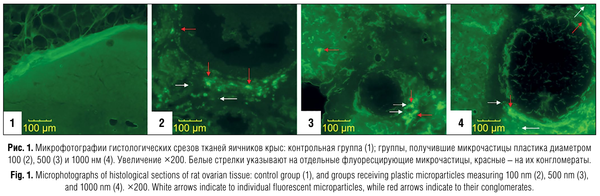
Fig. 2 (see on the insert) shows microphotographs of ovarian tissue sections from rats in the control group and groups exposed to MP, obtained in fluorescence mode at higher magnification. In the sections from the control group (2.1–2.3), predominantly stroma is visualised without signs of microparticle accumulation: there are no luminous rounded objects with distinct contours, nor are there any conglomerates of such objects. In the MP 100 group (2.4–2.6), round fluorescent objects, their clusters and individual conglomerates are observed in the brain parenchyma. In the sections of groups MP 500 (2.7–2.9) and MP 1000 (2.10–2.12), both single microparticles and their conglomerates are identified, localised predominantly in the cortical and cerebral layers; Figures 2.9 and 2.12 show the edges of the follicles with local clusters of microparticles around them.
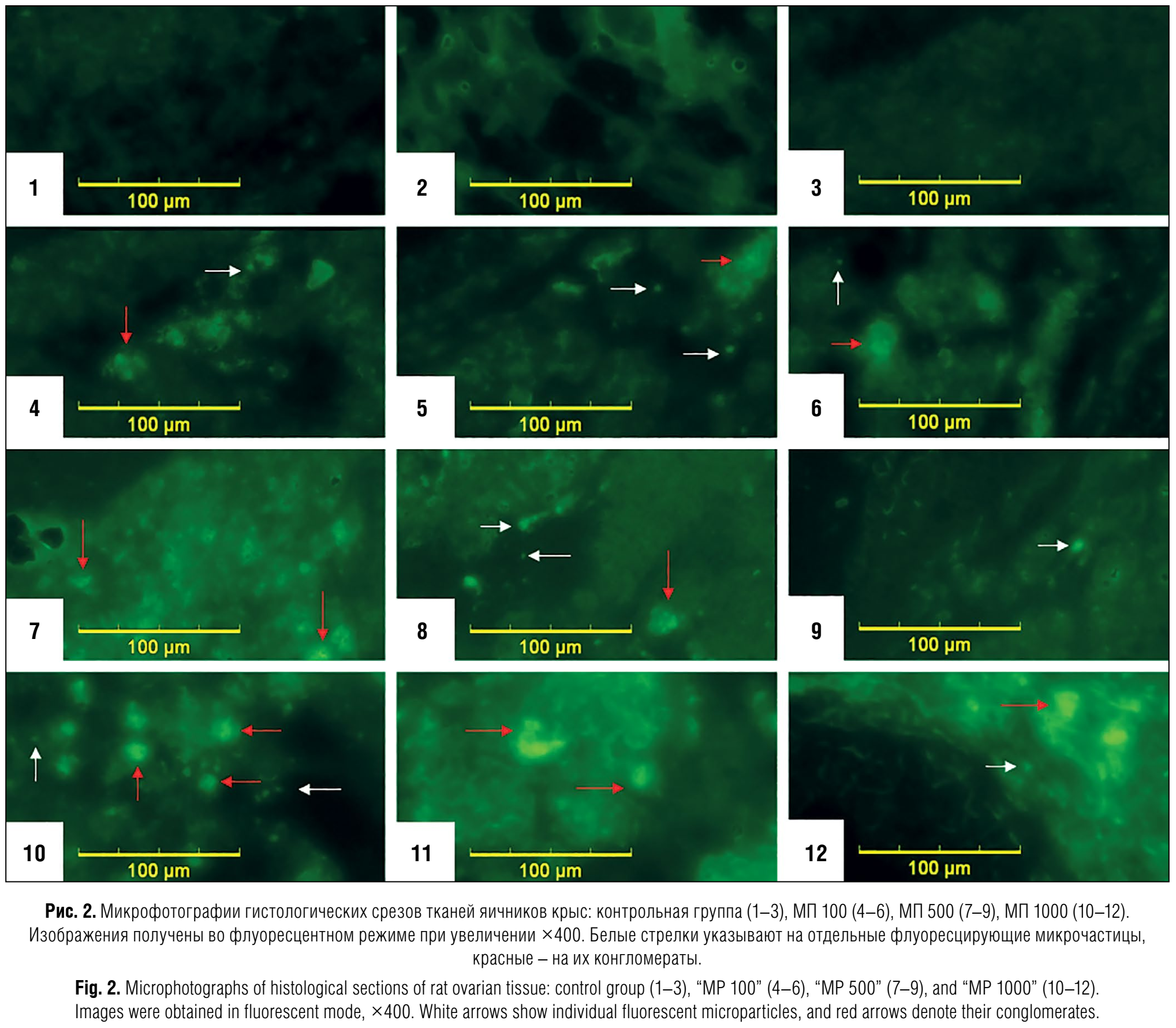
Fig. 3 presents the results of a quantitative assessment of the degree of accumulation and distribution of MP and its conglomerates in ovarian tissues. According to the analysis performed using a specially developed programme, isolated signals (0.42 ± 0.25 units) were recorded in the control group, likely due to background fluorescence and falling within the margin of error, as these animals were not exposed to MP particles. In the experimental groups, the number of fluorescent objects was significantly higher: 316.92 ± 44.03 units in the MP 100 group, 208.00 ± 73.08 units in the MP 500 group, and 979.1 ± 238.54 units in the MP 1000 group. The obtained values differed from the control values (р = 0.001 for the MP 100 and MP 1000 groups, р = 0.014 for the MP 500 group). Significant differences were also found between the MP 100 and MP 500 (р = 0.013), as well as between the MP 500 and MP 1000 groups (р = 0.007). The MP area in the tissues was assessed in pixels and revealed a similar trend: in the control group, the value was 1.42 ± 1.11 pixels, whereas in the experimental groups, the area increased significantly increased: to 9661.75 ± 1647.35 (MP 100), 5875.92 ± 1924.55 (MP 500) and 15,934.33 ± 3,488.31 pixels (MP 1000) respectively. In all cases, the differences from the control were statistically significant (р = 0.01). Significant differences were also observed between the MP 500 and MP 1000 groups (р = 0.012).
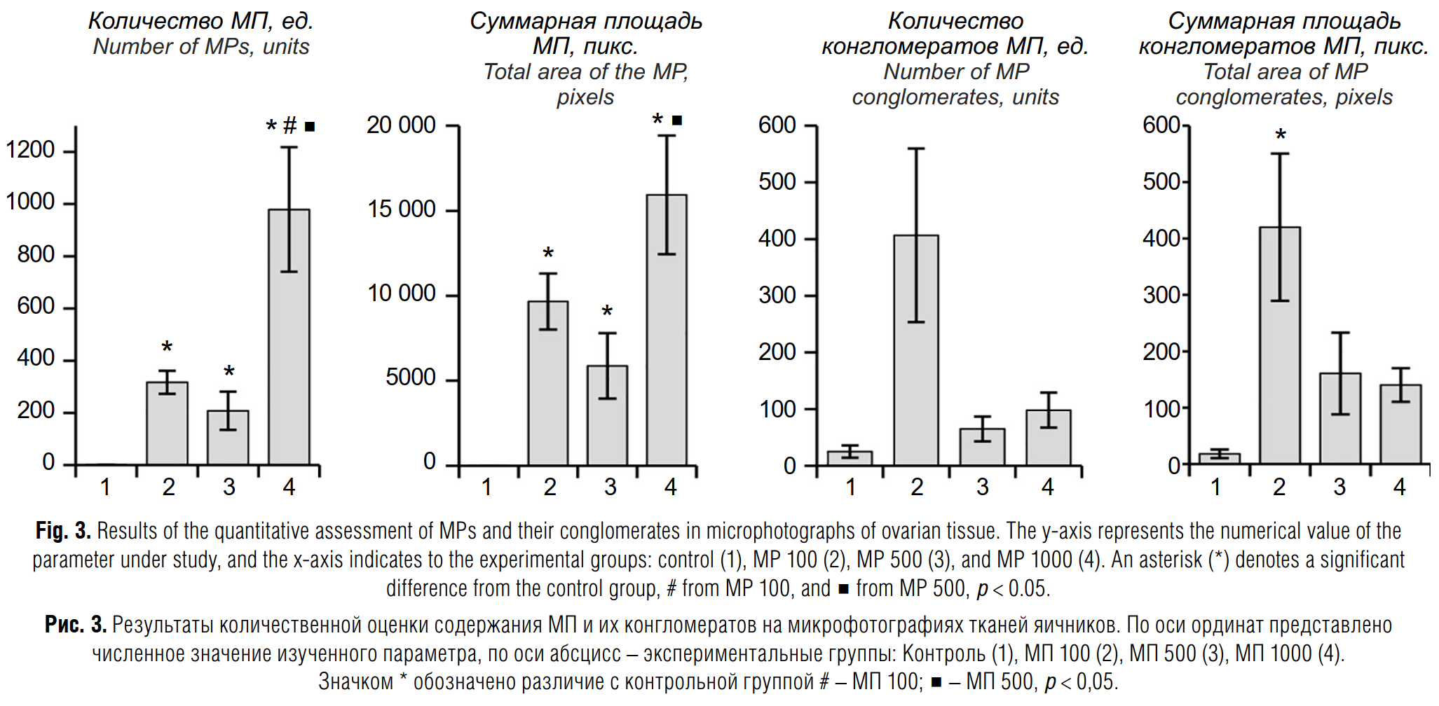
Analysis of the number of MP conglomerates revealed that in the control group, the number of suspected conglomerates was 25.08 ± 10.81 units, which may be due to misinterpretation of background fluorescence. At the same time, in the experimental groups, the values were significantly higher: 406.67 ± 153.18 units (MP 100), 65 ± 21.87 units (MP 500) and 98.17 ± 30.83 units (MP 1000). However, due to high variance, the intergroup differences did not reach statistical significance. Assessment of the area of conglomerates revealed that in the MP 1000 group (8400.58 ± 2611.56 pixels) and the MP 100 (2803.58 ± 595.51 pixels), the area was significantly higher than in the control (363.08 ± 154.13 pixels; the area may also be associated with misinterpretation of background fluorescence) at р = 0.010 and р = 0.001 respectively. In the MP 500 group, the area was also increased (3212.88 ± 1449.49 pixels), but the differences compared with the control did not reach statistical significance (р = 0.147); no significant differences were found between the experimental groups
Discussion
The ovaries possess a specific architecture combining a functionally active cortical zone, where the follicles are located, and a medulla with richly vascularised connective tissue. High vascularisation and intense hormonal metabolism make the ovaries potentially vulnerable to the accumulation and effects of xenobiotics, including MP [17]. Any changes in the structure or function of this organ may adversely affect reproductive function, the body’s hormonal status, and, likely, the quality of maturing follicles. Therefore, studying the penetration of MP into various zones of the ovary, the potential for the formation of intra-organ aggregates, and establishing size-dependent differences are of paramount importance when assessing potential risks of reproductive toxicity. It is known that direct contact between cells of a living system and MP can cause adverse effects at the cellular level [18].
The results obtained confirmed the ability of fluorescently labelled MP of 100, 500, and 1000 nm in size to penetrate the ovarian structure within a short period of time (5 h) following acute systemic administration (see Figs. 1, 2).
In a comparative analysis, we found the highest absolute number of recorded fluorescent objects in the 1000 nm MP group, which may be partly due to the better visibility and more intense fluorescence of larger particles. At the same time, the tendency observed in particles with a diameter of 100 nm to form extensive conglomerates suggests a possible tendency for smaller elements to aggregate within tissues. A similar effect of aggregation of smaller particles has been described in a number of experimental studies. Thus, in an experimental study with polystyrene microparticles (1, 3 and 6 µm) in fresh water, it was found that 84% of 1-µm particles form aggregates within 72 hours, whereas for 6-µm particles this figure is only 33% [19]. In another study, also devoted to the behaviour of MP in environmental conditions, it was shown that the critical coagulation concentration increases with increasing particle size, indicating a decrease in the stability of small particles [20]. Despite the lack of studies directly comparing the aggregation tendency of nanoparticles of different sizes in vivo it is known that in biological media, particles in the nano- and micrometre ranges exhibit pronounced size-dependent aggregation stability: as particle size decreases, they become less stable and more prone to agglomeration [21, 22]. In this study, we hypothesise that a similar mechanism may also apply to MP, although this hypothesis requires further experimental confirmation. Differences in the total surface area of individual MP also depend, as expected, on their diameter: the MP 1000 group exhibited the highest surface area per particle, yet the total surface area of their agglomerates was lower than in the MP 100 group. It is possible that larger microparticles, due to their size and, likely, differences in surface charge, are not prone to agglomerating into large clusters, but predominantly remain as isolated inclusions in tissues.
It is important to note that no additional immunofluorescent verification of the location of the vascular bed was performed in this study. Consequently, it cannot be ruled out that some of the visualised signals may have been located within the lumina of blood vessels, particularly given the extensive vascularisation of the ovarian parenchyma (see Figs. 1, 2). To distinguish more accurately between fluorescence in the tissue and the vascular bed, it will be necessary in future to use endothelial cell markers or to perform serial sections stained to identify blood vessels. It is also worth noting a factor such as the phase of the oestrous cycle, which potentially influences the permeability of tissue barriers and the metabolism of xenobiotics. In this series of experiments, we did not control for the phase of the cycle, which may have caused some variability in the results. However, statistically significant differences between the groups confirm that the effect of MP accumulation itself clearly outweighs the influence of hormonal fluctuations, at least in the context of acute exposure. Nevertheless, for an in-depth analysis of the effect of MP on reproductive function, it is important in future to include control of the cycle phase in the experimental design and to take into account data on the potential variability of vascular permeability during different periods.
A distinctive feature of this study was the use of the specialised Microplastic Detector software, which enables automated morphometric analysis and recognition of MP in fluorescent microphotographs. By systematically accounting for morphological criteria (roundness, size) and employing multi-stage thresholding, the software minimises operator error, ensuring data reproducibility even when working with a small number of samples. Unlike general-purpose tools, where the operator would have to manually set thresholds or configure macros for similar operations, Microplastic Detector automatically generates and consolidates results across a specified range of threshold values. Separate settings for conglomerates allow the specifics of each image set to be taken into account. Furthermore, the programme not only generates summary tables for subsequent statistical analysis but also saves visually annotated images, ensuring transparency and verifiability at all stages of processing. A key advantage of the programme is its standardised analysis protocol, which involves a one-time configuration of parameters, after which all images are processed identically: a feature particularly valuable when dealing with large datasets. In the future, expanding the functionality (including the use of machine learning methods and neural networks) will allow for improved accuracy in particle classification and the identification of more complex patterns of their distribution within tissues.
Thus, our data confirm that even brief exposure can lead to the accumulation of nanoparticles in ovarian tissues, and the nature of their localisation and the formation of aggregates depends on particle size. The observed differences in the aggregation of large and small particles indicate a complex interplay of physical and biological factors that determines the final distribution pattern of MP within the organ. Nevertheless, despite the growing body of data on the interaction of MP with living organisms, a comprehensive assessment of the impact of these particles on reproductive health requires further research focusing on hormonal regulation, the processes of ovulation and implantation, and possible genotoxic effects. An in-depth analysis will enable a more comprehensive understanding of the risks of MP reproductive toxicity and the development of effective methods to prevent its potential adverse effects.
Conclusion
It has been shown that plastic microparticles of 100, 500 and 1000 nm in size, when administered intracardially in a volume of 0.5 ml to outbred female rats, are detected in the ovaries. Thus, despite significant advances in understanding the mechanisms of penetration and accumulation of plastic microparticles, the specific effects on the reproductive system remain insufficiently studied.
One limitation of our study is the failure to account for the phase of the oestrous cycle in female rats, which may have influenced individual differences in the accumulation of plastic microparticles in the ovaries. Hormonal fluctuations during different phases of the cycle could alter tissue permeability and the accumulation of microplastics; therefore, future studies should take into account the animals’ hormonal status to allow for a more accurate interpretation of the data.
1. Mao X., Xu Y., Cheng Z., Yang Y., Guan Z., Jiang L., et al. The impact of microplastic pollution on ecological environment: a review. Front. Biosci. 2022; 27(2): 46. https://doi.org/10.31083/j.fbl2702046
2. Bora S.S., Gogoi R., Sharma M.R., Anshu B.M.P., Deka P., Bora J., et al. Microplastics and human health: unveiling the gut microbiome disruption and chronic disease risks. Front. Cell Infect. Microbiol. 2024; 14: 1492759. https://doi.org/10.3389/fcimb.2024.1492759
3. Jin H., Yang C., Jiang C., Li L., Pan M., Li D., et al. Evaluation of neurotoxicity in BALB/c mice following chronic exposure to polystyrene microplastics. Environ. Health Perspect. 2022; 130(10): 107002. https://doi.org/10.1289/EHP10255
4. Barua S., Mitragotri S. Challenges associated with Penetration of nanoparticles across cell and tissue barriers: a review of current status and future prospects. Nano Today. 2014; 9(2): 223–43. https://doi.org/10.1016/j.nantod.2014.04.008
5. Tao Z., Ghoroghchian P.P. Microparticle, nanoparticle, and stem cell-based oxygen carriers as advanced blood substitutes. Trends Biotechnol. 2014; 32(9): 466–73. https://doi.org/10.1016/j.tibtech.2014.05.001
6. Prüst M., Meijer J., Westerink R.H.S. The plastic brain: neurotoxicity of micro- and nanoplastics. Part Fibre Toxicol. 2020; 17(1): 24. https://doi.org/10.1186/s12989-020-00358-y
7. Liang B., Zhong Y., Huang Y., Lin X., Liu J., Lin L., et al. Underestimated health risks: Polystyrene micro- and nanoplastics jointly induce intestinal barrier dysfunction by ROS-mediated epithelial cell apoptosis. Part Fibre Toxicol. 2021; 18(1): 20. https://doi.org/10.1186/s12989-021-00414-1
8. Nie J.H., Shen Y., Roshdy M., Cheng X., Wang G., Yang X. Polystyrene nanoplastics exposure caused defective neural tube morphogenesis through caveolae-mediated endocytosis and faulty apoptosis. Nanotoxicology. 2021; 15(7): 885–904. https://doi.org/10.1080/17435390.2021.1930228
9. Yang D., Zhu J., Zhou X., Pan D., Nan S., Yin R., et al. Polystyrene micro- and nano-particle co-exposure injures fetal thalamus by inducing ROS-mediated cell apoptosis. Environ. Int. 2022; 166: 107362. https://doi.org/10.1016/j.envint.2022.107362
10. Repina E.F., Khmel A.O., Karimov D.D., Ryabova Yu.V., Karimov D.O., Kulagin E.A., et al. Distribution of 1000 nanometers plastic microparticles in vital organs after a single administration to laboratory animals. Mezhdunarodnyi zhurnal prikladnykh i fundamental’nykh issledovanii. 2025; (2): 40–4. https://doi.org/10.17513/mjpfi.13695 https://elibrary.ru/telmed (in Russian)
11. Repina E.F., Karimov D.O., Kudoyarov E.R., Karimov D.D., Akhmadeev A.R., Gizatullina A.A., et al. Preliminary assessment of the distribution of 500 nm plastic microparticles in rat organs after a single exposure. Meditsina truda i ekologiya cheloveka. 2025; (1): 113–25. https://doi.org/10.24412/2411-3794-2025-10109 https://elibrary.ru/bddeha (in Russian)
12. Hillery A.M., Jani P.U., Florence A.T. Comparative, quantitative study of lymphoid and non-lymphoid uptake of 60 nm polystyrene particles. J. Drug. Target. 1994; 2(2): 151–6. https://doi.org/10.3109/10611869409015904
13. Braakhuis H.M., Park M.V., Gosens I., De Jong W.H., Cassee F.R. Physicochemical characteristics of nanomaterials that affect pulmonary inflammation. Part Fibre Toxicol. 2014; 11: 18. https://doi.org/10.1186/1743-8977-11-18
14. Powell J.J., Faria N., Thomas-McKay E., Pele L.C. Origin and fate of dietary nanoparticles and microparticles in the gastrointestinal tract. J. Autoimmun. 2010; 34(3): J226–33. https://doi.org/10.1016/j.jaut.2009.11.006
15. Carr K.E., Smyth S.H., McCullough M.T., Morris J.F., Moyes S.M. Morphological aspects of interactions between microparticles and mammalian cells: intestinal uptake and onward movement. Prog. Histochem. Cytochem. 2012; 46(4): 185–252. https://doi.org/10.1016/j.proghi.2011.11.001
16. Romano J.P., Wolf M. Multiple testing of one-sided hypotheses: combining Bonferroni and the Bootstrap. In: Kreinovich V., Sriboonchitta S., Chakpitak N., eds. Predictive Econometrics and Big Data. TES 2018. Studies in Computational Intelligence, volume 753. Cham: Springer; 2018. https://doi.org/10.1007/978-3-319-70942-0_4
17. Ullah S., Ahmad S., Guo X., Ullah S., Ullah S., Nabi G., et al. A review of the endocrine disrupting effects of micro and nano plastic and their associated chemicals in mammals. Front. Endocrinol. (Lausanne). 2023; 13: 1084236. https://doi.org/10.3389/fendo.2022.1084236
18. Hwang J., Choi D., Han S., Jung S.Y., Choi J., Hong J. Potential toxicity of polystyrene microplastic particles. Sci. Rep. 2020; 10(1): 7391. https://doi.org/10.1038/s41598-020-64464-9
19. Guntram W., Drago C., Pawlak J. Biogenic aggregation of small microplastics alters their ingestion by a common freshwater micro-invertebrate. Front. Environ. Sci. 2020; 8: 574274. https://doi.org/10.3389/fenvs.2020.574274
20. Wang Y., Chen X. Aggregation behavior of polyethylene microplastics in the nearshore environment: The role of particle size, environmental condition and turbulent flow. Sci. Total. Environ. 2023; 901: 165941. https://doi.org/10.1016/j.scitotenv.2023.165941
21. Shrestha S., Wang B., Dutta P. Nanoparticle processing: Understanding and controlling aggregation. Adv. Colloid Interface Sci. 2020; 279: 102162. https://doi.org/10.1016/j.cis.2020.102162
22. Bélteky P., Rónavári A., Zakupszky D., Boka E., Igaz N., Szerencsés B., et al. Are smaller nanoparticles always better? Understanding the biological effect of size-dependent silver nanoparticle aggregation under biorelevant conditions. Int. J. Nanomedicine. 2021; 16: 3021–40. https://doi.org/10.2147/IJN.S304138
PhD (Medicine), head, Laboratory of toxicology, department of toxicology and genetics with an experimental laboratory animal clinic, Ufa research institute of occupational health and human ecology, Ufa, 450106, Russian Federation
e-mail: ryabovayuvl@yandex.ru
PhD (Biology), senior researcher, Laboratory of genetics, Department of toxicology and genetics with an experimental laboratory animal clinic, Ufa Research Institute of Occupational Health and Human Ecology, Ufa, 450106, Russian Federation
e-mail: ufniimt@mail.ru
PhD (Biology), head, Laboratory of Genetics, Department of toxicology and genetics with an experimental laboratory animal clinic, Ufa Research Institute of Occupational Health and Human Ecology, Ufa, 450106, Russian Federation
e-mail: Q.juk@yandex.ru
Junior researcher, Laboratory of Toxicology, Department of toxicology and genetics with an experimental laboratory animal clinic, Ufa Research Institute of Occupational Health and Human Ecology, Ufa, 450106, Russian Federation
e-mail: khmel.al01@gmail.com
PhD (Medicine), senior researcher, Laboratory of Toxicology, Department of toxicology and genetics with an experimental laboratory animal clinic, Ufa Research Institute of Occupational Health and Human Ecology, Ufa, 450106, Russian Federation
e-mail: e.f.repina@bk.ru
PhD (Medicine), head, Department of toxicology and genetics with an experimental laboratory animal clinic, Ufa Research Institute of Occupational Health and Human Ecology, Ufa, 450106, Russian Federation; N.A. Semashko National Research Institute of Public Health, Moscow, 105064, Russian Federation
e-mail: karimovdo@gmail.com
Junior researcher, Laboratory of Toxicology, Department of Toxicology and Genetics with an experimental laboratory animal clinic, Ufa Research Institute of Occupational Health and Human Ecology, Ufa, 450106, Russian Federation
e-mail: dgaar87@gmail.com
Researcher, Laboratory of Toxicology, Department of toxicology and genetics with an experimental laboratory animal clinic, Ufa Research Institute of Occupational Health and Human Ecology, Ufa, 450106, Russian Federation
e-mail: h-n-yu@yandex.ru
Junior researcher, Laboratory of Genetics, Department of toxicology and genetics with an experimental laboratory animal clinic, Ufa Research Institute of Occupational Health and Human Ecology, Ufa, 450106, Russian Federation
E-mail: e.kudoyarov@yandex.ru
Ryabova Yu.V., Mukhammadiyeva G.F., Valova Ya.V., Khmel A.O., Repina E.F., Karimov D.O., Akhmadeev A.R., Khusnutdinova N.Yu., Kudoyarov E.R. A comparative study of the accumulation of microplastics of different sizes in rat ovaries using an automated quantification algorithm. Hygiene and Sanitation. 2026;105(3):321-326. https://doi.org/10.47470/0016-9900-2026-105-3-321-326. EDN: pofgko
Мы в ВК:

 Email this article (Login required)
Email the author (Login required)
Email this article (Login required)
Email the author (Login required) 141014, The Moscow region, Mytishchi, 2, Semashko street
Federal Scientific Center of Hygiene named after F.F. Erisman
Phone: +7 916 885 28 02
e-mail: gigsan@list.ru